STEP 3: CT Scan

PA Diagnostic Flowchart CT Oct25
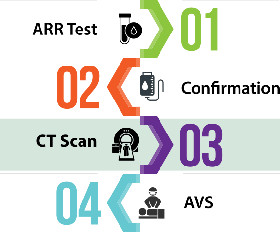
Once primary aldosteronism has been biochemically established by ARR and confirmatory testing, the next step in diagnosis consists in determining the subtype of the disease.

Although it only concerns about a third of patients, the form of primary aldosteronism for which treatment is the most effective is unilateral disease where an adenoma is the source of excess aldosterone.

In the third step of the diagnosis process, imaging of the abdomen is obtained by CT scan to assess whether there is an adenoma or the rare adrenal carcinoma on one of the adrenal glands.

What the Test Involves

A computed tomography scan (CT scan) is an imaging technique that produces cross-sectional views of the body with high resolution. Since a CT scan can distinguish tissues that differ in physical density by less than 1%, it is not only used to detect the presence of adrenal adenomas which can cause excess aldosterone, but also to assess whether the overall appearance of the adrenal glands is “bulky” and therefore indicative of bilateral disease.

To visualize the adrenal glands, the imaging technique is applied to the abdominal area, and involves the use of a contrast agent (usually iodine-based) to obtain functional information about the adrenal tissues and highlight structures such as blood vessels which would otherwise be difficult to distinguish from their surroundings.

You will lie on your back on a narrow table with your arms raised above your head. You will be asked to stay still to prevent blurred images, and may have to hold your breath for short periods of time. The contrast agent is usually given through a vein (IV) in the forearm, and within seconds of its administration you will likely feel a slight burning sensation, have a metallic taste in your mouth, and feel a warm flushing through your body. These feelings are all normal, and go away very rapidly.

How to Prepare for the Test and What to Do Afterwards

You may be asked not to eat or drink for 4 to 6 hours before the test. If you are diabetic and use metformin to treat it, you may also have to stop taking the drug before the test.

If you have an iodine allergy, your provider may give you antihistamines or steroids prior to undergoing the imaging test. Contrast agents rarely cause a life-threatening allergic response. If you experience difficulty breathing during the test, you should let the scanner operator know via the intercom and speakers.

Contrast agents are removed from the body by the kidneys. Hyperaldosteronism is associated with diabetes and impaired kidney performance. Some patients have reported a vulnerability to contrast agents. Not only must this be assessed prior to undergoing the CT scan, but you should also discuss the need for extra fluid intake after the test to help flush the iodine out of your body.

What Results Mean

Aldosterone-producing adenomas (APAs) may appear as small hypodense nodules — less dense than surrounding tissue — and are usually <2 cm in diameter.

Adrenal glands affected by idiopathic adrenal hyperplasia (IAH) may appear normal or may show nodular changes.

Aldosterone-producing adrenal carcinomas (i.e., cancer of the adrenal glands) are usually larger than 4 cm in diameter, and show suspicious characteristics.

“Incidentalomas” are more common the older we get, and not all adenomas cause excess aldosterone. Some APAs can be missed when they are very small.

What Radiologists Look for in Adrenal CT Scans

Magnifying glass and document

References